|
Legg-Calvé-Perthes
disease is a rare disease of the hip that afflicts approximately
1 in 1200 children. Of those children, only about one in
four are girls. About 5% of all diagnosed develop the disease
in both hips (bilaterally). Most of these children are very
active and often very athletic. The age of diagnosis is
usually between 2 and 12 years old, with the average age
of 6. Legg-Calve'-Perthes children tend to be of shorter
stature due to delayed bone age. The purpose of this pamphlet
is to provide you with more information to help you understand
this condition and some of the treatments.
What
is Legg-Perthes Disease?
Legg-Calvé-Perthes
disease (LCPD) is a form of osteonecrosis of the hip that
is found only in children. It is known by a few other names
such as ischemic necrosis of the hip, coxa plana, osteochondritis
and avascular necrosis of the femoral head. Most commonly
it is called Legg-Perthes disease, LCPD, or Perthes.
LCPD
is of unknown origin. It is known that bone death occurs
in the ball of the hip due to an interruption in blood flow.
As bone death occurs, the ball develops a fracture of the
supporting bone. This fracture signals the beginning of
bone reabsorption by the body. As bone is slowly absorbed,
it is replaced by new tissue and bone.
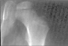 Initial
Phase Initial
Phase
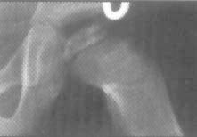 Reabsorption
Phase Reabsorption
Phase
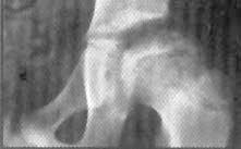 Reossification
Phase/Healed Reossification
Phase/Healed
Four
Stages of LCPD
-
Femoral head becomes more dense with possible fracture
of supporting bone;
-
Fragmentation and reabsorption of bone;
-
Reossification when new bone has regrown; and
-
Healing, when new bone reshapes.
Phase
I takes about 6-2 months, Phase 2 takes one year or more,
and Phase 3 and 4 may go on for many years.
Who
is at Risk?
There
is no specific cause known for LCPD, however, there are
some risk factors. Some of the factors identified as possible
links include children who are small for their age and are
extremely active. The disease is found more often in Asians,
Eskimos, and Whites, with a much lower incidence found in
Australian aboriginals, Native American, Polynesians and
Blacks. Exposure to secondhand smoke is correlated with
LCPD.
First
Symptoms
The
first symptoms characterized in LCPD are usually a limp
and perhaps pain in the hip, groin, or knee (known as a
referred pain). Often you will first notice limping during
your child's active play. They usually cannot tell you an
instance when they hurt themselves. They may not be able
to tell you exactly where they hurt, especially if the pain
is referred toward the knee area. They may not even experience
much pain. Other cases may not be diagnosed until some precipitating
event (fall, twisting injury) leads to an x-ray that uncovers
the previously undiagnosed Legg-Calve'-Perthes disease.
Diagnosis
Initial
diagnosis will require an x-ray, magnetic resonance imaging
(MRI) or bone scan. Other diagnostic measures may include
tests for limitation of abduction, a measurement of the
thigh to determine muscle atrophy, and tests to determine
the child's range of motion.
Extent
of Disease
It is
rare for a patient to have whole head involvement. However,
age can play an important role in the prognosis of the disease.
New bone growth typically reshapes better in younger children
and it may improve with growth.
There
are several different classifications used to determine
severity of disease and prognosis.
The
Catteral Classification
specifies four different groups to define radiographic
appearance during the period of greatest bone loss.
The
Salter-Thomson Classification
simplifies the Catteral Classifications by reducing them
down to two groups: Group A (Catteral I, II) which shows
that less than 50% of the ball is involved, and Group B
(Catteral Ill, IV) where more than 50% of the ball is involved.
Both classifications share the view that if less than 50%
of the ball is involved, the prognosis is good, while more
than 50% involvement indicates a potentially poor prognosis.
The
Herring Classification
studies the integrity of the lateral pillar of the ball.
In the Lateral Pillar Group A, there is no loss of height
in the lateral 1/3 of the head and little density change.
In Lateral Pillar Group B, there is lucency and loss of
height of less than 50% of the lateral height. Sometimes
the ball is beginning to extrude the socket. In Lateral
Pillar Group C, there is more than 50% loss of lateral height.
Many
doctors utilize these classifications as they provide an
accurate method of determining prognosis and help in determining
the appropriate form of treatment.
Prevention
There
is no known effective preventative measure.
TREATMENT
The
Goal
The
goal of treatment is four-fold:
I) to reduce hip irritability
2) restore and maintain hip mobility
3) to prevent the ball from extruding or collapsing
4) to regain a spherical femoral head
Types
of Treatment
Often
at the initial diagnosis, the physician may take a "wait
and see" approach to get a clearer picture of the progression
of the disease. As long as the patient's symptoms are mild,
the physician may only prescribe physical therapy exercises
to help maintain good range of motion. If the patient's
mobility changes, then the physician may prescribe either
non-surgical or surgical treatment.
Non-Surgical
Treatment
Non-surgical
treatments come in varying forms. Crutches are used for
non-weight bearing treatment for pain. Casts, traction,
and braces help return range of motion and mobility. Range
of motion exercises may be given to you by your physical
therapist to do with your child in the home.
Surgical
Treatment
Tenotomy
A
"Tenotomy" is a surgery that is performed to release
an atrophied muscle that has shortened due to limping. Once
released, a cast is applied allowing the muscle to regrow
to a more natural length. Cast time is about six to eight
weeks.
Osteotomy
There are different types of "osteotomies" (cutting
the bone to reposition it) and, depending on the need they
are performed at different stages of the disease. At times
with the softening of the ball, there is the possibility
of the ball slipping out of the socket. To protect it, a
femoral varus osteotomy, with or without rotation partially
redirects the ball into the socket.
Another
approach to surgically treating Legg-Calve'- Perthes is
to do an osteotomy above the hip socket. This allows the
surgeon to reposition the hip socket in such a way that
the femoral head will have less tendency to become deformed.
The shelf arthroplasty gives added coverage of the ball
from the top lip of the socket. Both the innominate and
the shelf arthroplasty help in reshaping.
LOOKING
TO THE FUTURE
Studies
on long-term results of LCPD indicate that the incidence
of late degenerative osteoarthritis is dependent on two
factors. If the ball reshapes well and fits well in the
socket, arthritis is usually not a concern. If the ball
does not reshape well, but the socket's shape still conforms
to the ball, the patient will tend to develop mild arthritis
in later adulthood. Patients whose femoral head does not
shape well and does not fit well in the socket usually develop
degenerative arthritis before the age of 50.
Although
Legg-Calvé-Perthes disease cannot be prevented, much
has been accomplished toward minimizing its effects. Research
and clinical studies continue to provide patients with better
long-term results.
The
National Osteonecrosis Foundation
Johns Hopkins University
P.O. Box 518
Jarrettsville, MD 21084
1-443-248-4889
www.nonf.org
|